MRI Scan Explained: What Happens During the Procedure?
By Dr. Mitusha Verma in Nanavati Max Advanced Institute of Radiation Oncology and Radiosurgery
Nov 14 , 2026

Recurring lower-back pain may sometimes require more detailed imaging, particularly when it starts travelling down a leg or is accompanied by tingling. If an examination suggests that the discs, nerves, or surrounding tissues need closer assessment, a doctor may recommend an MRI scan.
Knowing that an MRI has been recommended can raise questions about what happens inside the scanner. The machine can look enclosed, and the loud sounds and need to remain still may make the procedure seem unfamiliar at first. Understanding each step, from the safety checks to the final set of images, can make the experience more predictable.
Quick AnswerDuring an MRI scan, you lie on a movable table that slides into a large scanner. The machine uses a strong magnetic field and radio waves to create detailed images without using ionising radiation. The scan itself is painless, although you will hear loud knocking or tapping sounds and need to remain still while images are taken. Some MRI scans require intravenous contrast, while others do not. Most examinations are completed within an hour, although the exact duration depends on the type of scan. |
What Is an MRI Scan?
Magnetic resonance imaging, or MRI, is a non-invasive imaging test that produces detailed pictures of structures inside the body. It uses a strong magnetic field and radiofrequency waves rather than ionising radiation. MRI is particularly useful for examining soft tissues such as the brain, spinal cord, nerves, muscles, ligaments, joints and many internal organs. The type of imaging chosen depends on the condition being investigated.
Why Might Your Doctor Recommend an MRI?
An MRI may be recommended when detailed images could help clarify a diagnosis, assess an injury, or guide treatment.
It can be used to:
- Investigate Unexplained Symptoms: MRI can provide more detail when symptoms are not fully explained by an examination.
- Assess Injuries: It can show ligaments, muscles, cartilage, nerves, and other soft tissues.
- Evaluate Medical Conditions: MRI is widely used for neurological, spinal, cardiac, and abdominal conditions.
- Monitor Changes: Repeat scans may help assess a known condition or response to treatment.
- Support Treatment Planning: Detailed imaging may be required before certain procedures or surgeries.
The body part being examined determines the positioning and image sequences used during the scan.
Know about the Different Types of Radiology and Its Techniques
What Can an MRI Scan Examine?
MRI can be used across many areas of the body.
|
Type of MRI |
What It Can Help Examine |
|
Brain MRI |
Brain tissue, nerves, blood vessels, and neurological abnormalities |
|
Spine MRI |
Discs, spinal cord, nerves and surrounding structures |
|
Musculoskeletal MRI |
Ligaments, tendons, cartilage, muscles and joints |
|
Cardiac MRI |
Heart structure and function |
|
Abdominal MRI |
Liver, kidneys, pancreas and other abdominal organs |
|
Pelvic MRI |
Uterus, ovaries, prostate, bladder and surrounding structures |
|
Breast MRI |
Detailed breast imaging in selected situations |
How Should You Prepare for an MRI?
Most MRI scans require little preparation. However, safety screening is essential because the scanner produces a powerful magnetic field.
Before the scan:
- Remove jewellery, watches, hair clips and other metal objects.
- Leave phones, keys and other electronic items outside the MRI room.
- Follow any instructions about eating or drinking.
- Bring previous scans or reports when available.
- Tell the team about implants, medical devices or possible metal fragments.
- Mention pregnancy, kidney disease, dialysis or previous contrast reactions.
- Tell the team if enclosed spaces make you significantly anxious.
Doctor's NoteDo not assume that having an implant automatically means you cannot undergo MRI. Tell the radiology team exactly which device or implant you have so its MRI safety and any required scanning conditions can be checked beforehand. |
What Happens During an MRI Scan?
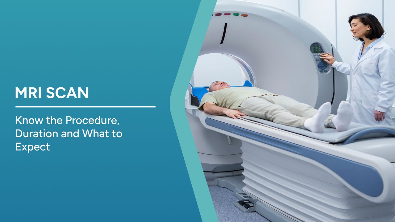
Once the safety checks are complete, you will lie on a padded table. The technologist positions you according to the area being examined and may use cushions or supports to help you remain comfortable. A specialised piece of equipment called a coil may be positioned close to the part being scanned. The table then moves into the scanner. How far you move inside depends on the body area being examined.
The technologist operates the scanner from an adjoining control room but can see and communicate with you throughout the examination.
During image acquisition, the scanner produces loud knocking, tapping, humming, or buzzing sounds. These noises are normal, and ear protection is generally provided. The scan consists of several image sequences with short pauses between them.
Why Do You Have to Stay Still During an MRI?
Movement can blur MRI images because each sequence takes time to record. If an image is affected by movement, that part of the examination may need to be repeated. For some abdominal or cardiac scans, you may also be asked to hold your breath briefly. If pain makes it difficult to remain in one position, tell the technologist before scanning begins.
Practical TipGet comfortable before the first sequence starts. If a support feels wrong or your existing pain makes the position difficult to maintain, tell the technologist immediately. Adjusting your position beforehand is easier than repeatedly moving during image acquisition. |
Will You Need Contrast During the MRI?
Not every MRI requires contrast. Some scans provide all the information needed without an injection, while others use contrast to make particular tissues, blood vessels, inflammation, or abnormalities easier to distinguish.
If contrast is needed, a small cannula is usually inserted into a vein in the hand or arm. Tell the medical team if you have kidney disease, are on dialysis, or have previously experienced a reaction to contrast.
How Long Does an MRI Scan Take?
The duration depends on the body part and type of examination. A relatively straightforward scan may take around 20 to 30 minutes, while more detailed examinations can take longer. Most MRI examinations are completed within an hour. Contrast administration, additional sequences or movement requiring repeated images can extend the scan.
What If You Are Claustrophobic?
If enclosed spaces make you anxious, tell the radiology team when scheduling the scan. Knowing what to expect and maintaining communication with the technologist can make the procedure easier.
At Nanavati Max Super Speciality Hospital, the Radiology Department has wide-bore 3 Tesla MRI capability. Its MAGNETOM Vida XT platform features a wider bore and faster image acquisition, which may improve comfort for people who feel anxious inside the scanner.
If claustrophobia is likely to prevent the examination from being completed, the medical team can discuss suitable options beforehand.
Know about the History of Radiology and Its Types
How Is an MRI Different From a CT Scan, X-Ray or Ultrasound?
Each imaging technique answers different clinical questions.
|
Test |
Key Difference |
|
MRI |
Uses magnetic fields and radio waves and provides detailed soft-tissue images |
|
CT Scan |
Uses X-rays to produce rapid cross-sectional images |
|
X-Ray |
Uses ionising radiation and is commonly used for bones and chest imaging |
|
Ultrasound |
Uses sound waves to create real-time images |
MRI offers detailed soft-tissue imaging without ionising radiation, while other techniques may be more appropriate depending on the condition and how quickly images are required.
Key Takeaway
- MRI uses a strong magnetic field and radio waves to produce detailed images without ionising radiation.
- Safety screening before the scan is important because metal and some medical devices require specific assessment.
- During the scan, you need to remain still while the machine produces normal knocking and tapping sounds.
- Contrast is used only when it is expected to provide additional diagnostic information.
- Tell the radiology team beforehand about implants, pregnancy, kidney disease, claustrophobia or difficulty remaining still.
Conclusion
An MRI scan is a non-invasive imaging test that can provide detailed views of the brain, spine, joints, nerves and many internal organs. The procedure involves safety screening, careful positioning, and a series of image sequences while you remain still inside the scanner. Loud noises are a normal part of image acquisition, and the technologist remains in communication with you throughout the examination. Contrast may be used when additional detail is required, but it is not necessary for every MRI.
Knowing what to expect can make a first MRI less uncertain, particularly if noise, claustrophobia or existing pain is a concern. Sharing information about implants, medical conditions, and anxiety before the scan also allows the radiology team to plan appropriately. Once the images are complete, a radiologist interprets them alongside the clinical question so the treating doctor can use the findings to guide further diagnosis or treatment.
Frequently Asked Questions
1. Is an MRI Scan Painful?
No. The MRI itself is painless. However, remaining in one position may become uncomfortable if you already have back, joint or other pain.
2. Does Your Whole Body Go Inside the MRI Machine?
Not always. How far you move into the scanner depends on the body part being examined. For some limb scans, the head may remain outside.
3. Can You Eat Before an MRI?
Most routine MRI scans do not require fasting. Some examinations have specific food or drink restrictions, so follow the instructions provided for your scan.
4. Is an MRI Safe During Pregnancy?
MRI does not use ionising radiation, but pregnancy should always be disclosed before the scan. The doctor and radiology team will decide whether the examination and any contrast are appropriate.
5. What Happens If You Move During an MRI?
Movement can blur the images and make them harder to interpret. If this happens, the affected sequence may need to be repeated.
Disclaimer
This article is intended for educational purposes only and should not replace professional medical advice, diagnosis or treatment. Follow the preparation and safety instructions provided for your individual MRI examination.

Written and Verified by: